|
Legends of the Echo images
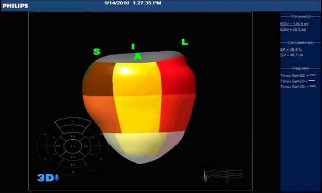
3D LV volume:
3-dimensional ECHO for LV volumetric analysis and calculation of ejection fraction.
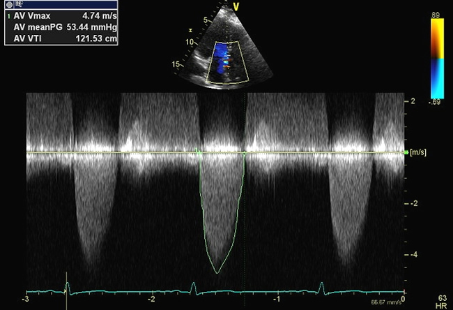
Aortic Valve CW gradient:
Continuous wave (CW) Doppler interrogation of trans-aortic valve gradient. A mean systolic gradient of 53 mmHg suggests significant aortic stenosis.
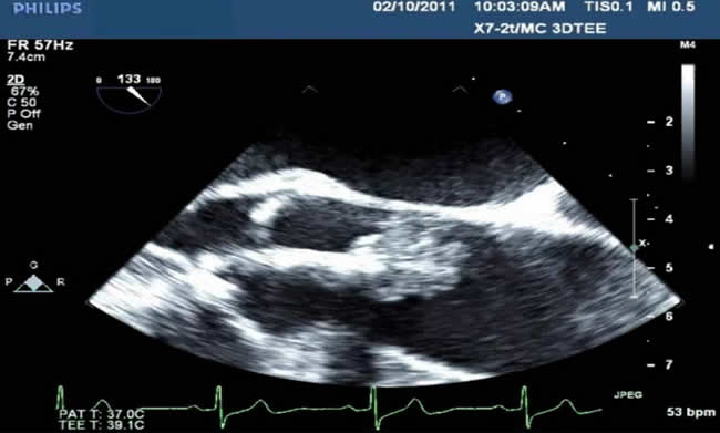
Aortic valve mass:
2D ECHO images demonstrating a huge papilloma-like mass attaching to the aortic valve.
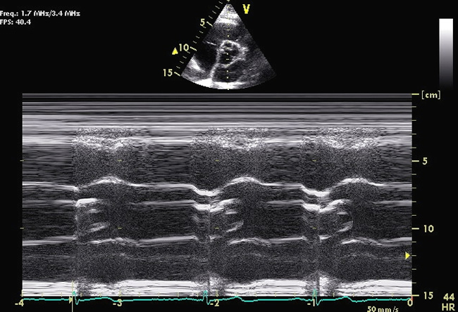
Bicuspid AV M mode:
M mode ECHO demonstrating eccentric aortic valve closure line of a bicuspid aortic valve during diastole.
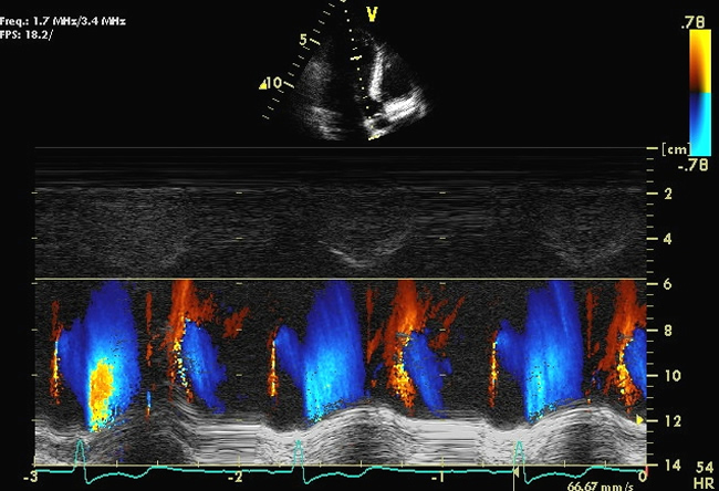
Color M mode:
M mode image with addition of color flow Doppler of LV cavity. Supplemental information for LV diastolic function could be obtained from the early diastolic flow propagation velocity.
|